Warning - today's video is not for the faint of heart.
We will be looking at the heart of a C. aceratus beating...but it is no longer in the icefish. You have been warned. But first, let's talk.
Let me tell you how that is possible for a beating heart to continue to contract outside of the body without being in a science fiction film or in an Edgar Allen Poe story. (And if you haven't read some Poe, let this serve as your inspiration) Our story begins back in 1882 with a doctor, a physiologist named Sidney Ringer.
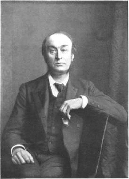
 Dr. Sydney Ringer. Picture courtesy of the Wellstone Trust.
Technically, it started before that. Remember, science is a never-ending quest to understand the world in which we live. What we discover today is built upon what has been discovered before us. As we began studying and learning more and more about the human body and physiology great strides were made in science. In the 1860s a German fellow, also a physician and a physiologist, named Carl Friedrich Wilhelm Ludwig (Let's just call him Dr. Ludwig) developed a way to study organs outside of the body - perfusion techniques. Perfusion - a great thing to know - especially if you are going to have open heart surgery. Technically perfusion means running a liquid over an organ. Most often that means blood, but it doesn't always. Organs that are going to be transplanted need to be in a preservative of some type while they are waiting their new home. Chemical compounds are run through the heart to make it stop when undergoing open heart surgery. That's called cardioplegia - cardo/heart, plegia/paralysis. In open heart surgery, the heart is stopped (much easier to work on than if it is constantly moving, don't you think?) Stopped? NO! You'll die. Well, yes you would if it was left like that, unless your doctor worked in warp speed and was done in seconds.
The main purpose of you heart is to circulate the blood which carries all sorts of important molecules and compounds into and out of the cells in your body. One of those being oxygen which is needed by your cells. It also needs to pick up the waste (like carbon dioxide, metabolic wastes, etc.) and get those to your lungs (CO2) and kidneys (metabolic and chemical waste - that's why urine is used for drug testing, by the way). Anyway, your blood needs to constantly circulate. So if your heart isn't doing it - modern technology has a machine to do it. And the person who does this in the operating room is called - any guesses? - the perfusionist! Great.
Back to Ringer. He was a doctor and he taught (hooray for teachers!!) medicine also. He was British. He was developing a way to keep organs and tissues 'alive', so to speak, longer outside of the body. The story has it that his lab tech was accidentally using tap water instead of distilled water. This tap water had calcium in it (calcium is involved in muscle contractions) and it worked better. Voila! Scientific history in the making by accident. Happens more than you might imagine. This was in 1882 and he was working on frog hearts.
Today, Ringer's solution is commonplace in hospitals and labs. The trick is that it is a solution that contains ingredients (compounds for those of you that have had chemistry, or should I say remember the chemistry you have had) that are mixed together in the same concentration that naturally occur in your body fluids. Ringer's solution has sodium chloride, potassium chloride, calcium chloride, and sodium bicarbonate.
Almost movie time. Stay with me. One last question to answer. Why do we use Ringer's solution when dissecting our fish? We need to get accurate weights of their organs. An organ with blood in it obviously weighs more than an organ without blood. Think about wet clothes and how much more they weigh when they are drenched with water. So to get that blood out, we put it in Ringer's solution so the heart pumps it out.
In the interest of scientific education, I filmed an icefish heart to show you. It's a great example of how the heart works as well.
http://
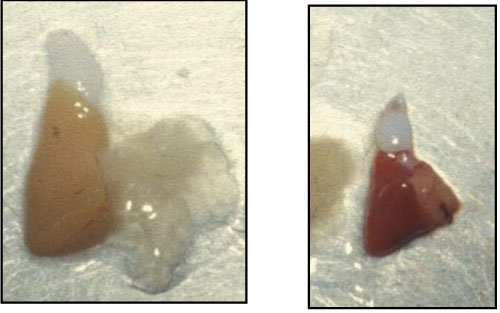
For those of you that want further description - the gel-like part of the heart is the atrium. This is where the blood enters heart. The tan, meaty looking part is the ventricle. The white thing on the top is the bulbus arteriosis. In this area the blood pressure is lowered before it is pumped back into the gills. If you watch closely you can see the atrium contract and then the ventricle contract. There is a lot of interesting stuff to say about their hearts. Let me put a plug in for my webinar on all of this. It is archived on this site and you can listen or just look at the pictures.
 Compare the hearts of the icefish and the coriiceps. Icefish (C. aceratus) is on the left. N. coriiceps is on the right. These fish are about the same size yet the icefish heart is much larger.
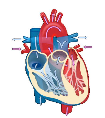
Our hearts look very different but we have similar structures. Humans have two atria and two ventricles. Basically, our heart is two pumps.
 Here is a human heart. The arrows show the flow of blood. Pink arrows mean blood rich with oxygen. Blue arrows signify blood that is oxygen poor. Whether it is oxygen rich blood headed to the body or oxygen poor blood headed to the lungs to stock up on O2, they both enter the atrium. It just depends on which side of the heart it enters. Right side takes in oxygen poor blood. Left side sends out the oxygen rich blood to the body.
