Why study phytoplankton?
Why study these microscopic floating plants? Because despite their tiny size, they are critical to much of life on Earth. They are the foundation of the bountiful marine food web, produce oxygen for the air we breath and take in harmful carbon dioxide. That's why scientists are so interested in learning more about them.
To collect phytoplankton the crew and science team launch a CTD Rosette. This rosette can carry several oceanographic tools including the Niskin sea water collection bottles. On the R/V Sikuliaq the CTD rosette is operated by a winch system that lowers the rosette below the ocean surface to collect water samples. Within those water samples are the phytoplankton!
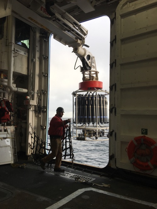 Ethan Roth the Marine Science Tech on the R/V Sikuliaq draws a safety gate as the CTD Rosette with the Niskin bottles gets ready to go below the surface.
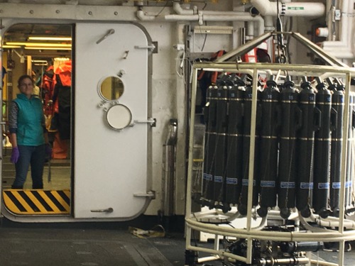 Dr. Lowry watches over the Niskin bottles with her water samples as the large door that leads from the Baltic Room to the sea closes. August 29, 2017. Photo by Lisa Seff.
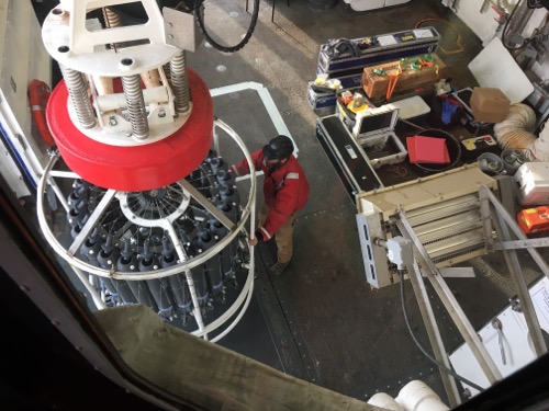 View of the CTD Rosette with the Niskin bottles from the winch operating room above the Baltic Room. August 29, 2017. Photo by Lisa Seff.
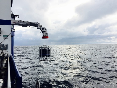 The CTD Rosette along with the Niskin bottles heads below the surface of the Beaufort Sea. August 30th, 2017.
The Niskin bottles are open as the rosette is lowered into the ocean and then they are snapped shut at predetermined depths by a computer! The sea water and plankton are then divided into different sample bottles for a variety of tests and procedures. Dr. Lowry, a postdoctoral scholar at the Woods Hole Oceanographic Institution, uses the FlowCAM as both a microscope and a camera to capture photos of the phytoplankton for use in future analysis.
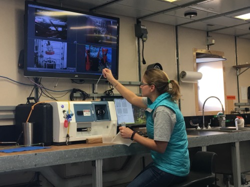 Dr. Kate Lowry calibrates and sets up the FlowCAM with an underway sea water sample. She's hoping the green laser light will work as it's key to capturing great photos of phytoplankton.
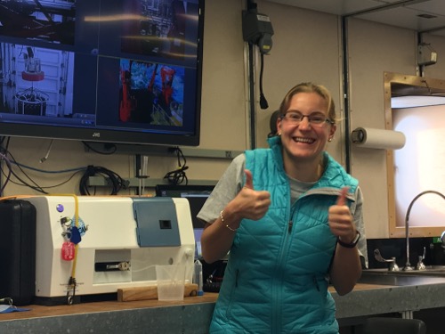 What does it take to make a very happy research scientist? A fully functioning green laser light in the FlowCAM! August 27, 2017.
These are some of the first images we collected, from the ship's underway water intake system, of microscopic phytoplankton using the imaging FlowCAM. The cells seen in the photo below are 100 times their actual size!
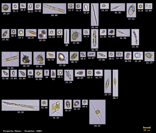 One of the first phytoplankton photographs using the FlowCAM on the R/V Sikuliaq! Photo by Dr. Lowry. August 29, 2017.
After the water is retrieved from the Niskin bottles, the sea water is filtered to keep only the smallest microscopic phytoplankton.
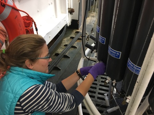 Dr. Lowry collects water from a Niskin bottle. August 30, 2017. Photo by Lisa Seff.
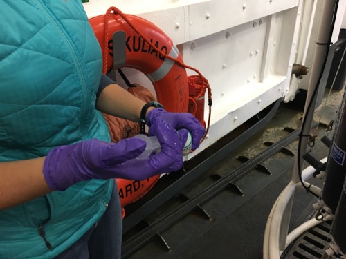 Dr. Lowry filters the sea water to remove larger particles and organisms. August 30, 2017. Photo by Lisa Seff.
It's important to filter out larger plankton or other particles because anything too large might clog the tubing that the water flows through in the FlowCAM. Once the sample is ready Dr. Lowry runs the filtered water samples through the FlowCAM which uses a green laser to trigger the phytoplankton to fluoresce, this then triggers the camera to take an image! The images are saved automatically so Dr. Lowry and other research scientists can analyze them later to determine what species of phytoplankton were found at that test site.
Chlorophyll Analysis
In addition to her photographing phytoplankton Dr. Lowry also uses water she collects from the Niskin bottles to analyze levels of chlorophyll in the water. This process allows her to determine the approximate biomass of the plankton at a specific location. It’s is a multi-step, time consuming process so she’s asked me to help her in the analytical lab. I’m very excited because it’s something I always wanted to learn! Dr. Lowry made a step by step protocol as every sample must be treated the same way, so we don’t introduce any other variables that could compromise the data. While working with the samples we keep a log of everything for the same reason and make sure we note if we do anything different with any of the samples. Then, if a data set appears out of line we can easily check back in our notes to see if anything different was going on with that particular sample. I’m finding the process to be a lot of fun, very relaxing and similar to a mindfulness exercise, as when you're doing the work you really need to focus and be in the moment or it’s easy to make a mistake.
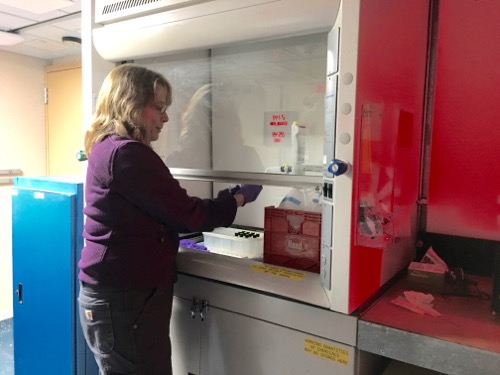 Science teacher Lisa Seff adds hydrochloric acid to filtered chlorophyll samples. August 27, 2017. Photo by Dr. Kate Lowry.
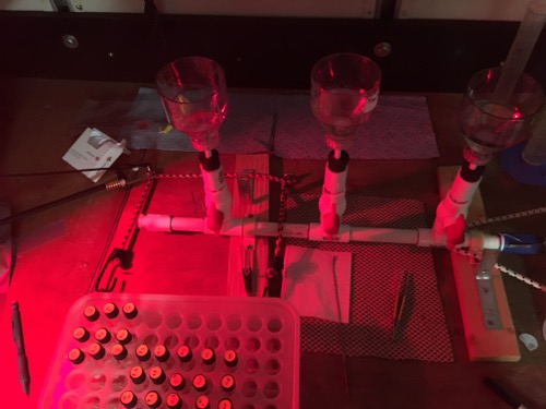 When working with the chlorophyll it's important to use a red light so the chlorophyll doesn't degrade. August 29, 2017. Photo by Lisa Seff.
Through the Porthole! Arctic artwork from Springs School Students, Anvil City Science Academy and East Hampton community members!
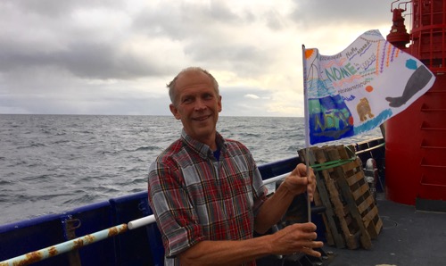 Arctic artwork flag from students at the Anvil City Science Academy in Nome! August 30, 207. Photo by Lisa Seff.
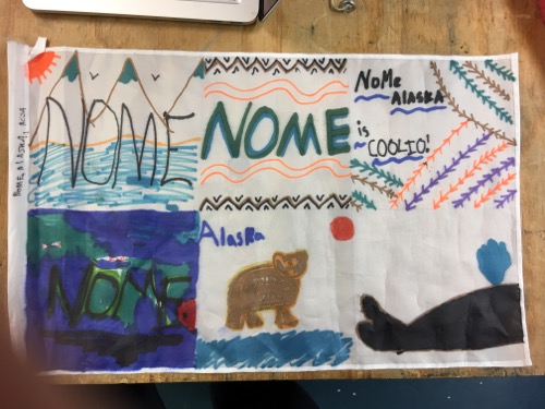 Arctic artwork flag from students at the Anvil City Science Academy in Nome! August 30, 207. Photo by Lisa Seff.
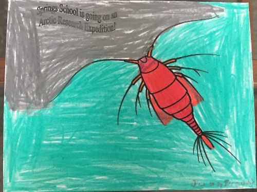 Arctic organism artwork from Springs School student Juan! Photo by Lisa Seff. August 2017.
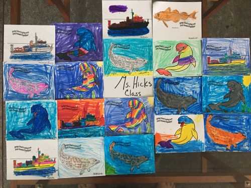 Arctic artwork by Mrs. Hick's class 2016/2017. August 2017. Photo by Springs School PolarTREC educator Lisa Seff.
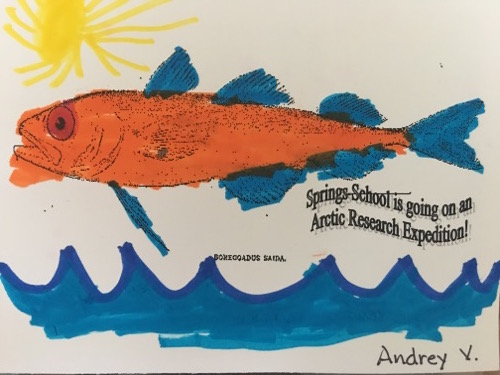 Arctic artwork by Andrey. August 2017. Photo by Springs School PolarTREC educator Lisa Seff.
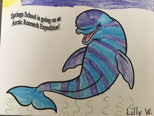 Arctic artwork by Springs School student Lilly W. August 2017. Photo by Springs School PolarTREC educator Lisa Seff.
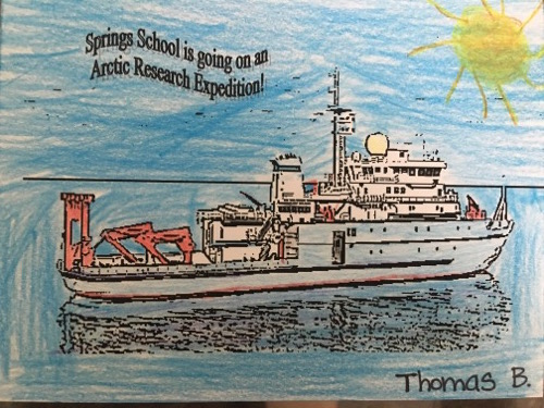 Arctic artwork by Thomas B.! August 2017. Photo by Springs School PolarTREC educator Lisa Seff.
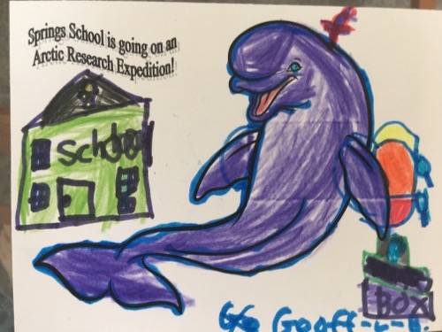 Arctic organism artwork from Springs School student Geoff! Photo by Lisa Seff. August 2017.
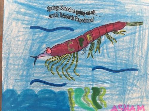 Artwork by Springs School student Asham.. June 2017. Photo by Lisa Seff. August 2017.


Comments
Add new comment